
The Dental Chair Is Only the Beginning: The Future of Connected Digital Dentistry
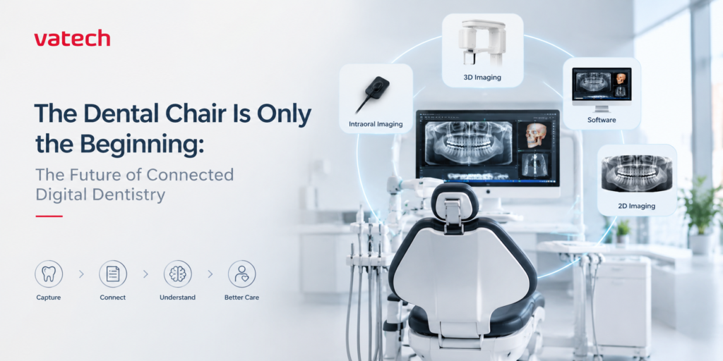
Why the modern dental practice needs more than individual devices
For generations, the dental chair has been the centre of the dental practice.
It is where the patient sits.
It is where examination happens.
It is where treatment is delivered.
But modern dentistry no longer begins and ends at the chair.
Before a procedure, there is patient history, examination and imaging. During diagnosis, there may be intraoral radiographs, panoramic imaging or CBCT. Treatment planning may involve multiple sources of clinical information. After diagnosis, those same images become part of the conversation between dentist, patient, specialist and laboratory.
The dental chair is still where dentistry happens. But increasingly, the information around it shapes how dentistry happens.
And that changes the way dental technology should be evaluated.
The problem isn’t a lack of technology
Walk into a modern dental practice and you may find an impressive collection of digital equipment.
An intraoral sensor.
An OPG.
A CBCT.
Perhaps an intraoral scanner.
Different software platforms.
Different viewers.
Different systems storing different pieces of information.
Each device may perform its intended function very well.
Yet the overall workflow can still feel disconnected.
The dentist may have excellent images but spend time moving between systems.
The team may have digital equipment but still rely on manual steps to organise information.
The patient may see several different images without understanding how they relate to one another.
This is the paradox of digital dentistry:
A practice can have more technology and still have a fragmented workflow.
That is why the conversation around digital dentistry is gradually moving beyond simply acquiring more devices.
The more important question is becoming:
How well does the technology work within the clinical workflow?
Digital does not automatically mean connected
There is an important distinction between being digital and being connected.
Digital means information is captured, stored or processed electronically.
Connected means that information can become useful across different stages of the clinical journey.
Consider an implant case.
The patient may begin with a clinical examination and an intraoral radiograph. A panoramic image may provide a broader view. CBCT may then be required for three-dimensional assessment and treatment planning.
The value is not in having three separate images.
The value is in how those images contribute to one clinical decision.
The same principle applies to orthodontics, endodontics, oral surgery, prosthodontics and restorative dentistry.
Technology becomes more valuable when it helps the dentist move from:
Image → Information → Understanding → Decision.
The FDI World Dental Federation’s 2025 policy statement on digital dentistry similarly emphasizes evidence-based, patient-centred and outcome-driven use of digital technologies, alongside interoperability and continued professional judgment.
The dentist doesn’t need more information. The dentist needs useful information.
This is where technology companies have an important responsibility.
It is easy to add features.
It is much harder to make technology genuinely useful.
A CBCT system should not simply produce a three-dimensional image. It should help answer a clinical question.
An OPG should not simply generate a panoramic image. It should become part of diagnosis and treatment planning.
An intraoral sensor should not simply capture an X-ray. It should make acquisition efficient and the resulting image clinically useful.
Software should not become another layer of complexity. It should help clinicians access, interpret and work with the information they have already captured.
The objective is not more technology.
The objective is better clinical utility.
This is where Vatech’s approach matters
For a dentist evaluating imaging technology, the decision should go beyond comparing one specification with another.
It should begin with a more fundamental question:
What kind of imaging ecosystem do I want to build for my practice?
This is where Vatech becomes relevant.
Vatech’s dental portfolio spans 2D imaging, 3D imaging, intraoral imaging and software, giving dentists options across different stages of their imaging journey.
From intraoral imaging for everyday diagnostic needs to panoramic systems and CBCT for more advanced clinical applications, the portfolio is built around the different imaging requirements of modern dentistry.
The significance of this breadth is not simply that there are multiple products to choose from.
It is that a dentist can think about imaging as a long-term technology strategy, rather than as a series of unrelated purchases.
Start with the clinical question, not the machine
Suppose a dentist is considering a CBCT.
The first question should not be:
“Which CBCT has the largest FOV?”
It should be:
“What clinical cases do I want this system to help me manage?”
Implant planning?
Endodontics?
Impacted teeth?
Orthodontics?
Or a combination of applications?
The appropriate FOV, resolution, workflow, software and system configuration should follow from that clinical requirement.
The same principle applies to every imaging investment.
A practice should not buy technology simply because it is newer.
It should buy technology because it solves a meaningful clinical or workflow problem.
That is why Vatech’s broad imaging portfolio matters.
It allows practices to evaluate their imaging requirements across intraoral, 2D and 3D technologies rather than looking at every imaging need in isolation.
Image quality is important. What you do with the image is equally important.
For a long time, conversations about dental imaging focused heavily on image quality.
And rightly so.
But image quality is only the beginning.
Once an image has been captured, the dentist needs to work with it.
It needs to be viewed.
It needs to be interpreted.
It may need to be compared with other clinical information.
It may need to be explained to the patient.
It may need to support treatment planning.
And sometimes it needs to be shared with another professional.
That is why imaging software and viewing environments are becoming increasingly important parts of the digital dental workflow.
Vatech’s software portfolio includes image-processing and machine-learning technologies designed to support dental imaging workflows.
The larger idea is simple:
The value of an imaging system should not stop when the exposure ends.
The patient is part of the digital workflow too
There is another benefit of better imaging that is often underestimated:
Communication.
A patient may not understand a clinical explanation involving bone levels, root anatomy, an impacted tooth or a periapical lesion.
But showing the patient the relevant image can change the conversation.
Instead of asking the patient to imagine the problem, the dentist can show it.
Instead of simply recommending treatment, the dentist can explain the clinical reasoning behind it.
Technology therefore has a role not only in diagnosis, but also in making diagnosis understandable.
The image is no longer only something the dentist sees.
It can become part of what the patient understands.
Why choose Vatech?
This brings us to the question that every dentist should ask before making a technology investment:
Why this manufacturer?
The answer should never be simply a brand name.
It should come down to what the technology can contribute to the practice.
For a dentist considering Vatech, there are several factors worth evaluating.
A broad imaging portfolio
Vatech offers dental imaging solutions across intraoral, 2D and 3D imaging, allowing practices to select technology according to their clinical requirements.
Clinical relevance
Imaging technology should serve clinical decision-making rather than simply add another piece of equipment to the practice.
Imaging supported by software
The workflow does not end with image acquisition. Viewing, processing and working with diagnostic information are equally important parts of the experience.
A technology roadmap
A dental practice rarely stays exactly the same.
The cases change.
The team grows.
The procedures evolve.
The imaging requirements become more sophisticated.
Technology therefore needs to be considered as part of the practice’s future—not only its present.
The smarter investment is not always the most expensive one
There is a temptation in digital dentistry to equate advancement with complexity.
The newest technology.
The largest specifications.
The most features.
But the best technology investment is not necessarily the one with the longest specification sheet.
It is the one that answers the practice’s clinical needs while fitting the way the team actually works.
Before investing, dentists should ask:
What clinical problems am I trying to solve?
Which imaging modality do I actually need?
How will the system fit into my existing workflow?
What software and viewing tools will my team use?
How easily can the information be accessed, interpreted and communicated?
What training and technical support are available in my market?
Can this investment continue to make sense as my practice grows?
These questions move the conversation away from simply buying equipment.
They move it towards building a practice.
The dental chair is only the beginning
The future of dentistry will not be defined by how many digital devices a practice owns.
It will be defined by how effectively those technologies support the clinical journey.
From the first image to diagnosis.
From diagnosis to treatment planning.
From treatment planning to patient communication.
From one clinical decision to the next.
The dental chair remains at the centre.
But the technology surrounding it increasingly determines how efficiently information moves through the practice and how effectively that information can be turned into clinical action.
That is why choosing imaging technology is no longer simply a purchase decision.
It is a workflow decision.
It is a clinical decision.
And increasingly, it is a long-term practice decision.
Vatech’s portfolio across intraoral, 2D and 3D dental imaging gives dentists the opportunity to approach that decision with more than a single machine in mind.
Because the question is no longer:
“Which machine should I buy?”
It is:
“What kind of dental practice am I building—and what should my technology help me achieve?”
And perhaps that is where the modern dental practice truly begins.
- Published in Uncategorized
How Dental Lasers Work: Clinical Applications, Safety, and Evidence for Indian Dental Practice
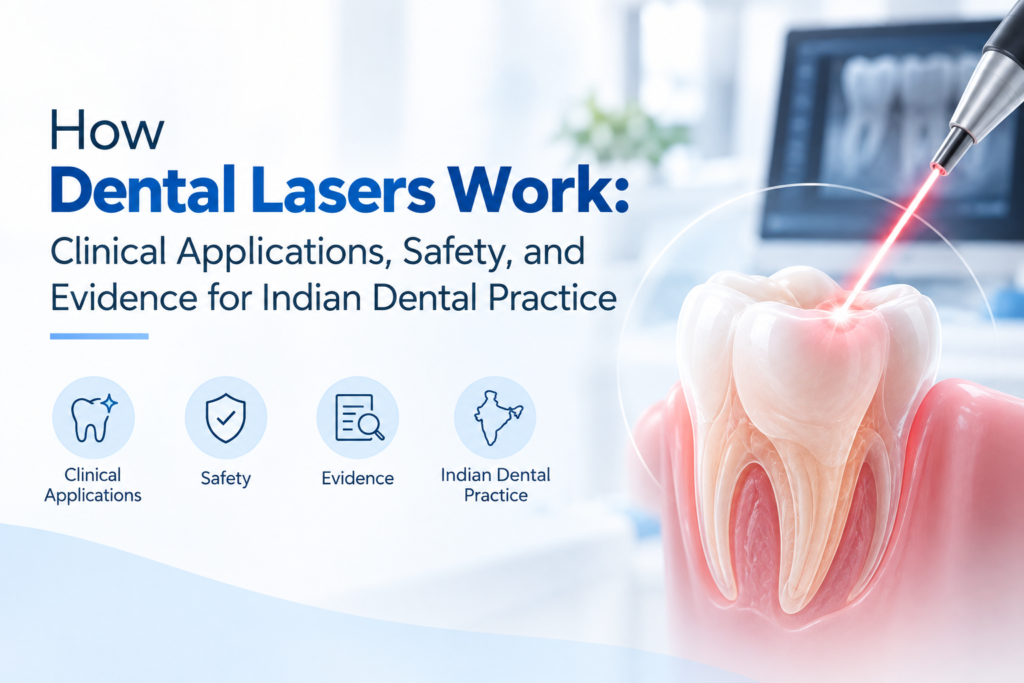
Dental lasers have become an increasingly discussed technology in modern dentistry. From soft-tissue procedures and periodontal therapy to selected endodontic applications, lasers can provide dentists with an additional tool for treatment where controlled light energy can interact with oral tissues.
But what exactly happens when a laser is used in dentistry? Is dental laser treatment suitable for every procedure? And what does current evidence actually say about its clinical benefits?
The answer depends on the laser wavelength, power, exposure time, tissue type and clinical indication. Dental lasers are not a replacement for conventional treatment in every case. Instead, they can serve as an adjunct or alternative for selected procedures when used according to appropriate protocols and training.
What Are Dental Lasers and How Do They Work?
A dental laser is a device that produces a concentrated beam of light at a specific wavelength. When this energy reaches oral tissue, it can produce different biological effects depending on the wavelength and how the laser is operated.
Soft-tissue dental lasers are commonly used because certain wavelengths interact strongly with pigments and water-containing tissues. This allows dentists to perform controlled procedures such as soft-tissue incision, contouring, coagulation and selected periodontal applications.
The clinical effect is influenced by several parameters, including:
- Laser wavelength
- Power or energy settings
- Continuous or pulsed operation
- Exposure time
- Distance and movement of the fiber
- Tissue characteristics
This is why dental laser safety depends not simply on owning a laser but on using the correct settings and technique for the intended procedure.
The Basic Physics: What a Dental Laser Actually Does
A laser produces a narrow beam of light that is monochromatic (a single wavelength) and coherent (the light waves travel in phase). What happens when that beam hits tissue depends almost entirely on the wavelength and on which molecules in the tissue absorb it.
Dental lasers work through a handful of interactions:
- Absorption — the target chromophore (water, haemoglobin, melanin, or hydroxyapatite) absorbs the light energy and converts it to heat or a photochemical effect.
- Photothermal (ablative) effect — enough energy absorption vaporises or cuts soft tissue; this is the basis of most surgical dental laser work.
- Photochemical / photobiomodulation effect — at low power, light triggers cellular signalling (mitochondrial activity, cytokine modulation) without significant heating, which is the basis of low-level laser therapy (LLLT).
- Photosterilisation — light-activated antimicrobial effects, either through direct thermal bacterial kill or via photodynamic therapy with a photosensitising dye.
Two wavelengths dominate the diode category used in general dental practice:
- 980 nm (near-infrared) — well absorbed by haemoglobin and water, making it effective for soft-tissue cutting and coagulation in contact mode, with a documented trans-analgesic effect that reduces the need for anaesthesia in many soft-tissue procedures.
- 650 nm (red light) — poorly absorbed by haemoglobin, penetrates tissue more deeply, and is the wavelength typically used for photobiomodulation and photodynamic applications rather than cutting.
Devices that combine both wavelengths (sometimes marketed as “double coherence”) are built to cover ablative and biomodulation work from one console, switching presets by procedure type.
Reading the Evidence Critically
A few patterns show up repeatedly across recent reviews, and they’re worth keeping in mind when evaluating any single study or manufacturer claim:
- Parameter variability is a real problem. Power settings, pulse durations, and fibre/tip diameters differ widely between studies, which makes pooling results and drawing firm conclusions difficult. Several 2025–2026 reviews explicitly call for standardised reporting of laser parameters.
- Study sizes are often small, and blinding a laser-versus-scalpel comparison to the operator is inherently difficult, which introduces bias risk that reviewers using tools like Cochrane RoB or ROBINS-I consistently flag.
- “Laser” is not one intervention. Diode, Er:YAG, Nd:YAG, and CO2 lasers behave very differently in tissue. Evidence for one wavelength or laser type should not be assumed to transfer to another.
- Comfort and bleeding outcomes are better supported than long-term healing or survival outcomes. Most positive findings relate to intraoperative and short-term postoperative measures; longer-term comparative data is comparatively sparse.
Dental Laser Applications in Dentistry
One of the reasons laser dentistry has attracted attention is its versatility. Different laser systems and wavelengths have different indications, so dentists should evaluate the technology based on the procedures they actually perform.
1. Periodontal Treatment
One of the most studied applications of Diode lasers in dentistry is periodontal therapy.
A diode laser may be used as an adjunct to conventional periodontal treatment in selected cases. Research has investigated its use alongside scaling and root planing, with some studies reporting additional clinical benefits while others have found limited or inconsistent differences.
A systematic review and meta-analysis of 30 randomized controlled trials found additional clinical benefits from adjunctive diode laser treatment in certain periodontal protocols, although outcomes varied according to application method and treatment regimen.
This means lasers should be viewed as a clinical adjunct rather than a universal replacement for conventional periodontal therapy.
2. Endodontics and Root Canal Treatment
Another important area of dental laser applications is endodontics.
Laser technology has been investigated for root canal disinfection, microbial reduction, irrigation assistance and other endodontic procedures. Recent literature reviews have identified several potential applications, including root canal disinfection, access preparation, regenerative procedures and postoperative pain management.
A 2026 systematic review and meta-analysis also found that diode laser-assisted approaches may provide advantages in bacterial reduction and early postoperative pain in some endodontic protocols, while noting that differences in laser parameters and study methods make the evidence difficult to standardize.
For Indian dental practices, this makes laser technology particularly relevant for clinicians interested in incorporating adjunctive technologies into endodontic workflows.
3. Soft-Tissue Procedures
Soft-tissue applications are another common area where laser dentistry can be useful.
Depending on the device and indication, dental lasers may be used for procedures involving gingival tissue and other soft tissues. The controlled delivery of laser energy can support cutting, tissue contouring and coagulation.
The clinical objective is not simply to use a laser instead of a conventional instrument. The advantage comes from having another tool that can be selected when its tissue interaction is appropriate for the procedure.
4. Periodontal and Peri-Implant Applications
Laser technology has also been studied in periodontal and peri-implant therapy. However, the level of evidence varies considerably between applications.
Current research continues to investigate how different wavelengths, protocols and treatment combinations affect clinical outcomes. A recent systematic review of periodontal laser surgery found promising but variable results across diode laser studies, highlighting the importance of standardized protocols and appropriate case selection.
Therefore, dentists should evaluate evidence for the specific procedure and laser protocol, rather than assuming that every laser application provides the same clinical benefit.
What Are the Benefits of Dental Lasers?
When appropriately indicated, dental lasers can offer several workflow and clinical advantages. These may include precise soft-tissue management, controlled energy delivery and the ability to perform selected procedures with a different tissue-interaction profile compared with conventional instruments.
For dentists considering a dental diode laser, practical benefits may include:
- Compact technology suitable for chairside use
- Controlled delivery through a small fiber
- Applications across selected soft-tissue and periodontal procedures
- Integration into minimally invasive treatment workflows
- Potential support for patient comfort in appropriate procedures
However, these benefits depend heavily on the specific device, clinical indication and operator technique.
Dental Laser Safety: What Dentists Need to Know
Safety should be one of the first considerations when introducing a dental laser into clinical practice.
Laser light can cause eye injury because the eye can focus concentrated laser energy onto the retina. The FDA classifies lasers according to their potential hazard, with higher classes representing greater potential risk when used improperly.
For dental practices, safety should include appropriate protective eyewear, controlled access to the treatment area, correct device settings, staff training and following the manufacturer’s instructions for use.
Most importantly, dentists should not select laser parameters simply by copying settings from another procedure or device. Wavelength, power, pulse characteristics and exposure time all matter.
Choosing a Dental Laser for an Indian Dental Practice
For dentists in India considering laser technology, the decision should go beyond the machine’s specifications.
Think about the procedures you perform most frequently and whether the laser’s wavelength and indications match those requirements. Training, ergonomics, fiber availability, service support and ease of integration into the existing workflow are also important.
Before investing in a dental laser, consider:
- Intended clinical applications
- Laser wavelength and operating modes
- Available power and control settings
- Fiber and accessory options
- Training requirements
- Safety features
- Service and technical support
- Manufacturer documentation and clinical indications
The right system is ultimately the one that fits the clinical workflow, training level and treatment needs of the practice.
The Future of Laser Dentistry
Dental lasers are continuing to evolve as research explores their applications across periodontics, endodontics, soft-tissue procedures and other areas of dentistry.
For Indian dental practices, the opportunity is not simply to adopt a new technology because it is modern. The greater value comes from understanding where laser technology can genuinely improve or complement an existing clinical workflow.
As evidence develops, dentists will be better positioned to use lasers selectively, safely and according to specific clinical indications.
Conclusion
Dental lasers are becoming an important technology in modern dental practice, particularly for selected soft-tissue, periodontal and endodontic applications. Their effectiveness depends on the wavelength, treatment parameters, clinical indication and operator expertise.
Current evidence suggests that lasers can provide useful adjunctive benefits in certain procedures, but results are not uniform across every application. For that reason, dentists should combine scientific evidence, clinical experience and patient-specific requirements when deciding where to use laser technology.
For practices considering a dental diode laser, the focus should be on practical clinical applications, appropriate training, safety and long-term workflow value.
Explore modern dental laser technology and discover how laser-assisted workflows can complement your clinical practice.
- Published in Uncategorized
Apex Locator: How It Improves Accuracy in Root Canal Treatment
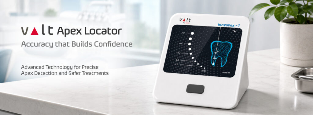
Root canal treatment is one of the most technique-sensitive procedures in dentistry. One of the most important steps in successful endodontic treatment is determining the correct working length of the root canal. Accurate working length helps dentists clean, shape, and obturate the canal effectively while reducing the risk of over-instrumentation or inadequate cleaning.
This is where an apex locator becomes an important part of modern endodontic practice.
An apex locator is an electronic dental device designed to help determine the position of the apical constriction and estimate the working length of a root canal. By providing real-time information during treatment, an electronic apex locator can support greater precision and efficiency during root canal procedures.
What Is an Apex Locator?
An apex locator is an electronic device used in endodontics to determine the approximate position of the root canal apex. It works by measuring electrical characteristics within the root canal and helps the dentist identify the location of the file relative to the apical region.
Traditionally, dentists have relied on radiographs, tactile sensation, and clinical experience to estimate working length. While radiographs remain an important diagnostic tool, an apex locator provides additional information during the procedure.
Modern electronic apex locators are designed to provide continuous or near real-time measurements, helping dentists make more informed decisions while preparing the root canal.
Why Is Working Length Important in Root Canal Treatment?
Working length determination is a critical step in root canal treatment. The objective is to establish the length at which cleaning and shaping should be performed while maintaining control of the apical region.
If the working length is too short, parts of the root canal may remain inadequately cleaned or disinfected. On the other hand, excessive instrumentation beyond the desired endpoint can potentially cause unnecessary irritation to the surrounding tissues.
Accurate working length determination therefore plays an important role in predictable endodontic treatment.
An apex locator can help dentists assess working length during the procedure and complement radiographic evaluation.
How Does an Electronic Apex Locator Work?
An electronic apex locator uses electrical measurements to estimate the position of the file inside the root canal.
During the procedure, the dentist places an endodontic file into the canal and connects it to the apex locator. The device measures changes in electrical characteristics as the file approaches the apical region.
The information is then displayed on the device, allowing the dentist to monitor the file position.
The exact technology varies between apex locator models. Modern devices may use advanced measurement methods designed to improve performance under different canal conditions.
However, an apex locator should be considered a clinical aid rather than a replacement for the dentist’s judgement, radiographs, and appropriate endodontic techniques.
Key Benefits of Using an Apex Locator
1. Improved Working Length Determination
One of the biggest advantages of an apex locator is its ability to assist with working length determination. Having an electronic measurement during treatment can help dentists establish an appropriate endpoint for canal preparation.
2. Real-Time Measurement
Unlike methods that provide information only after an image is taken, an electronic apex locator can provide measurements while the procedure is being performed.
This can help dentists monitor file position throughout the treatment.
3. Supports More Predictable Endodontic Treatment
Accurate canal preparation is an important part of successful root canal treatment. By assisting with working length determination, an apex locator can support a more controlled endodontic workflow.
4. Complements Radiographic Evaluation
An apex locator does not necessarily replace dental X-rays. Instead, it can complement radiographic assessment by providing electronic information about the file position inside the canal.
Using multiple sources of clinical information can help dentists make better treatment decisions.
5. Helps Improve Treatment Efficiency
Modern dental practices increasingly focus on efficient workflows without compromising clinical quality. An electronic apex locator can help streamline the working length determination process and reduce dependence on repeated estimation.
Apex Locator vs. Traditional Working Length Methods
Radiographs have been widely used for working length estimation in endodontics. However, a two-dimensional radiographic image may not always provide complete information about the three-dimensional anatomy of the root canal system.
An apex locator provides another source of information by electronically assessing the position of the file within the canal.
For this reason, the question is not necessarily whether dentists should choose an apex locator or radiographs. In many clinical situations, these methods can work together.
The dentist can use clinical examination, radiographs, electronic measurements, and clinical judgement to establish an appropriate working length.
When Should Dentists Consider Using an Apex Locator?
An apex locator can be useful across a wide range of root canal procedures. It may be particularly helpful when accurate working length determination is challenging because of complex root canal anatomy.
Curved canals, overlapping anatomical structures, and limitations of conventional radiographic interpretation can make endodontic procedures more demanding.
Having an electronic measurement tool available can give dentists additional information during canal preparation.
As with any dental equipment, proper use, calibration or device-specific protocols, and clinical judgement are important for obtaining reliable results.
How to Choose the Right Dental Apex Locator
When selecting a dental apex locator, dentists should consider several factors rather than focusing on a single feature.
Look for a device that offers:
- Accurate and consistent measurements
- A clear and easy-to-read display
- Simple operation
- Reliable performance in different clinical conditions
- Convenient file connection and handling
- A compact design suitable for everyday practice
- Easy maintenance and cleaning
- Reliable technical and after-sales support
The right apex locator should fit naturally into the dentist’s existing endodontic workflow.
Conclusion
An apex locator has become an important technology in modern endodontics because it assists dentists in determining root canal working length with electronic measurements.
By providing real-time information about file position, an electronic apex locator can complement radiographs and clinical assessment, helping dentists achieve a more controlled and efficient root canal procedure.
Ultimately, successful root canal treatment depends on several factors, including diagnosis, case selection, access preparation, cleaning and shaping, irrigation, obturation, and clinical expertise. An apex locator is one of the tools that can support dentists in achieving greater precision during this process.
For dentists looking to improve their endodontic workflow, understanding how an apex locator works and how it can complement conventional methods is an important step toward more predictable root canal treatment.
- Published in Uncategorized
CBCT vs OPG: What Is the Difference and Which One Does Your Dental Clinic Need?
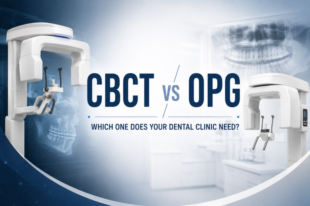
CBCT vs OPG: What Is the Difference and Which One Does Your Dental Clinic Need?
Dental imaging has completely changed the way dentists diagnose oral conditions and plan treatments. Today, dental clinics have access to technologies that can provide everything from a quick panoramic overview to highly detailed three-dimensional images. But with several imaging options available, one question is becoming increasingly common: CBCT vs OPG—which one is better for a dental clinic?
The answer is not as simple as choosing one technology over the other. CBCT and OPG are designed for different clinical requirements. An OPG provides a broad two-dimensional view of the teeth and jaws, while a CBCT machine produces three-dimensional images that allow dentists to examine anatomical structures from different perspectives.
Understanding the difference between CBCT and OPG is important for dentists planning to upgrade their imaging setup, particularly when considering factors such as clinical applications, image quality, workflow, radiation exposure, and long-term practice requirements.
What Is an OPG in Dentistry?
An OPG (Orthopantomogram), commonly known as a panoramic X-ray, provides a broad 2D image of the teeth, upper and lower jaws, and surrounding structures in a single scan.
For many dental clinics, an OPG is a practical imaging solution because it provides a complete panoramic overview quickly and conveniently. Instead of taking multiple individual dental X-rays, the dentist can obtain a single image covering a large area of the oral and maxillofacial region.
OPG imaging is commonly used during general dental examinations, orthodontic assessments, wisdom tooth evaluations, and preliminary treatment planning. It can also help dentists identify certain abnormalities involving the teeth and jaw.
Some common applications include:
- General dental assessment
- Wisdom tooth evaluation
- Orthodontic assessment
- Preliminary implant assessment
- Evaluation of developing teeth
- General jaw examination
The biggest strength of an OPG machine is its ability to provide a quick and comprehensive 2D overview. When the clinical question does not require detailed three-dimensional information, an OPG can be an efficient choice.
What Is a CBCT Machine?
A CBCT (Cone Beam Computed Tomography) machine takes dental imaging to another level by producing three-dimensional images of the scanned area.
Instead of presenting the anatomy as a single 2D image, CBCT creates a volumetric dataset that can be viewed in different planes. This gives dentists a much better understanding of the relationship between teeth, bone, nerves, sinuses, and other anatomical structures.
This additional information can become particularly valuable in complex cases where a conventional panoramic image may not provide enough detail.
CBCT in dentistry is commonly used for implant planning, complex endodontic cases, impacted teeth, oral surgery, orthodontic treatment planning, and evaluation of certain anatomical or pathological conditions.
For example, when planning an implant, knowing only the apparent height of the bone on a 2D image may not be enough. The dentist may also need to understand the available bone width and the position of important anatomical structures. This is where 3D imaging can provide valuable additional information.
CBCT vs OPG: What Is the Difference?
The most important difference between CBCT vs OPG is the type of image each technology produces.
An OPG creates a two-dimensional panoramic image, whereas CBCT produces a three-dimensional representation of the scanned area.
This difference affects how the images can be used during diagnosis and treatment planning. An OPG is excellent when the dentist needs a broad overview, while CBCT becomes more useful when the clinical situation requires detailed information about depth, bone dimensions, or anatomical relationships.
| Feature | OPG | CBCT |
|---|---|---|
| Image type | 2D | 3D |
| Main purpose | Panoramic overview | Detailed 3D assessment |
| Depth information | Limited | Available |
| Implant planning | Preliminary | Detailed |
| Impacted teeth | Initial assessment | 3D evaluation |
| Complex cases | Limited information | More detailed information |
| Image viewing | Panoramic view | Multiple planes and 3D views |
However, this doesn’t mean that CBCT is simply a replacement for OPG. Both technologies have their own place in modern dental imaging.
The right choice depends on what the dentist needs to see and what clinical decision needs to be made.
CBCT vs OPG for Dental Implants
Dental implant planning is one of the clearest examples of where the difference between CBCT and OPG becomes important.
An OPG can provide an excellent initial overview of the patient’s teeth and jaws. It can help the dentist evaluate the general condition of the area and identify potential concerns. However, implant placement is a three-dimensional procedure, and the dentist may need more detailed information before finalizing the treatment plan.
A CBCT scan for implant planning can provide information about bone dimensions and the location of important anatomical structures. This can help dentists evaluate the implant site from multiple perspectives rather than relying only on a 2D image.
CBCT can be particularly useful for assessing:
- Bone height and width
- Implant positioning
- Nerve canal location
- Sinus anatomy
- Complex anatomical structures
- Areas requiring additional surgical planning
This doesn’t mean every implant patient automatically needs CBCT. The decision should be based on the individual case and whether the additional 3D information is necessary for diagnosis and treatment planning.
CBCT vs OPG for Wisdom Teeth
Wisdom tooth assessment is another common application of panoramic imaging.
An OPG can show the position of an impacted wisdom tooth and provide a useful overview of its relationship with surrounding structures. For straightforward cases, this information may be sufficient for treatment planning.
However, some impacted teeth have a more complicated relationship with surrounding anatomical structures. If the dentist needs additional three-dimensional information, a CBCT examination may provide a more detailed view.
This is particularly relevant when there is concern about the relationship between an impacted mandibular third molar and the inferior alveolar nerve canal.
The important consideration is not simply whether CBCT provides more information, but whether that additional information will affect the diagnosis, treatment plan, or management of the patient.
When Is an OPG the Better Choice?
An OPG can be an excellent choice when the dentist needs a broad overview of the teeth and jaws without requiring detailed three-dimensional information.
For routine examinations and several common dental applications, the simplicity and speed of panoramic imaging can make an OPG a practical part of the clinic’s diagnostic workflow.
An OPG may be particularly useful for:
- Routine dental assessment
- Orthodontic evaluation
- Wisdom tooth assessment
- General jaw examination
- Initial treatment planning
- Assessment of developing dentition
For clinics handling a high volume of routine cases, having an OPG machine can make panoramic imaging a quick and convenient part of everyday diagnosis.
When Is CBCT the Better Choice?
A CBCT machine becomes particularly valuable when the dentist needs information that cannot be adequately obtained from a conventional 2D image.
For example, complex implant cases may require information about bone dimensions and anatomical structures. Similarly, complicated endodontic cases may benefit from additional information about root anatomy, while oral surgery may require a better understanding of the three-dimensional anatomy of the treatment area.
CBCT can therefore be useful when three-dimensional information is important for:
Implant planning: Understanding the available bone and surrounding anatomical structures.
Complex endodontics: Evaluating complicated root anatomy or cases where conventional imaging does not provide sufficient information.
Impacted teeth: Understanding the three-dimensional position of an impacted tooth.
Oral surgery: Supporting planning for procedures involving complex anatomy.
Selected orthodontic cases: Providing additional 3D information when clinically indicated.
The key principle is simple: CBCT should be used when the clinical question requires three-dimensional information.
Is CBCT Better Than OPG?
This is probably the most common question people ask when comparing CBCT vs OPG.
The answer is: not universally.
CBCT provides significantly more three-dimensional information, but that does not mean it should automatically be used for every dental patient. If a panoramic image provides all the information required for a particular clinical decision, an OPG may be completely appropriate.
Professional dental guidance emphasizes selecting radiographic examinations according to the individual patient’s clinical needs rather than using advanced imaging routinely without a specific diagnostic reason.
In other words, the goal should not be to choose the technology with the most advanced specifications. The goal should be to choose the right imaging technology for the clinical question.
CBCT vs OPG: What About Radiation?
Radiation exposure is an important factor when selecting dental imaging technology.
In general, CBCT examinations can involve more radiation than conventional dental radiography, although the actual dose varies significantly depending on factors such as the equipment, field of view, exposure settings, and scanning protocol.
This is why responsible imaging selection is important. Dentists should consider whether the additional information provided by a CBCT scan is clinically justified.
Modern imaging systems also incorporate dose optimization technologies and different scanning protocols, allowing imaging parameters to be selected according to the examination.
For patients and dental clinics, the focus should therefore be on appropriate imaging, optimized exposure, and clinically justified examinations rather than simply choosing one technology based on radiation alone.
Should a Dental Clinic Have Both CBCT and OPG?
For many growing dental practices, having access to both technologies can provide greater flexibility.
An OPG can handle cases where a broad panoramic overview is sufficient, while CBCT can be used when detailed 3D information is required. This allows the dentist to select the appropriate imaging method according to the patient’s clinical needs.
Some modern dental imaging systems combine panoramic and CBCT capabilities within a single unit. This type of setup can be particularly useful for multidisciplinary practices that handle general dentistry alongside implants, orthodontics, endodontics, and oral surgery.
Instead of asking “CBCT or OPG?”, a better question for a growing clinic may be:
“Which imaging capabilities does my practice need today, and where do I want my practice to be in the next five to ten years?”
That approach makes the equipment investment more strategic.
What Should You Consider Before Buying a CBCT or OPG Machine?
Buying dental imaging equipment is a long-term investment, so the decision should go beyond the initial purchase price.
Start by looking at your clinical workload and treatment mix. A general dental practice may have different imaging requirements compared with an implant-focused or multidisciplinary clinic.
Image quality is another important consideration. For CBCT, factors such as voxel size, field of view (FOV), image resolution, and dose optimization should be evaluated according to your clinical requirements.
Software and workflow also matter. A powerful imaging system should be supported by software that dentists and staff can use efficiently. Features for image viewing, measurement, treatment planning, and data management can make a significant difference to everyday usability.
Finally, don’t overlook after-sales service and technical support. Dental imaging equipment is a long-term investment, and installation, training, maintenance, warranty, and service availability can have a major impact on the overall ownership experience.
Before making a purchase, consider:
- Your most common clinical procedures
- Required imaging area and FOV
- Image quality requirements
- Radiation optimization
- Software capabilities
- Ease of use
- Installation and training
- Warranty and after-sales support
- Future expansion of your practice
CBCT vs OPG: Which One Does Your Dental Clinic Need?
There is no universal answer to whether your clinic needs a CBCT machine or an OPG machine. The right choice depends on the type of dentistry you perform and the kind of diagnostic information you regularly require.
If your practice mainly needs panoramic imaging for routine diagnosis, orthodontic assessment, wisdom teeth, and general treatment planning, an OPG can be a highly useful and practical solution.
If your clinic regularly performs implant planning, complex endodontics, oral surgery, or other procedures that require detailed three-dimensional information, CBCT can provide significant additional clinical value.
For a growing or multidisciplinary dental practice, having access to both can create a more complete dental imaging workflow.
The most important thing is not simply buying the most advanced equipment. It is choosing technology that matches your clinical requirements, improves your workflow, supports better treatment planning, and remains useful as your practice grows.
Conclusion
The comparison between CBCT vs OPG is ultimately not about finding one technology that is better for every situation. Both play important roles in modern dental imaging.
An OPG provides a quick and comprehensive 2D panoramic view, making it useful for many routine dental and orthodontic applications. CBCT, on the other hand, provides 3D dental imaging, allowing dentists to examine bone, teeth, nerves, sinuses, and other anatomical structures in greater detail when clinically required.
For dental clinics planning to upgrade their imaging capabilities, the decision should be based on clinical requirements rather than technology alone. Consider the types of cases you handle, the imaging information you need, your workflow, software requirements, service support, and the future direction of your practice.
As digital dentistry continues to evolve, having the right imaging technology can help dentists make more informed treatment decisions and create a more efficient, modern clinical workflow.
The right imaging system isn’t necessarily the one with the most features—it’s the one that gives your practice the right information when you need it.
- Published in Uncategorized
7 Essential Dental Equipment Every Modern Dental Clinic Needs in 2026
The dental industry is evolving faster than ever before. Patients today expect accurate diagnosis, minimally invasive treatment, faster appointments, and a comfortable clinical experience. At the same time, dentists are looking for technologies that improve efficiency, reduce chair time, and deliver predictable treatment outcomes. This shift has made investing in the essential dental equipment a necessity rather than a luxury.
A modern dental clinic is no longer defined by its location or interiors—it is defined by the technology it uses. From digital imaging systems and intraoral scanners to dental lasers and AI-powered software, advanced equipment enables dentists to diagnose conditions earlier, plan treatments with greater precision, and provide a superior patient experience.
In this guide, we’ll explore the 10 essential dental equipment every modern dental clinic should consider in 2026 and explain how each technology contributes to clinical excellence and practice growth.
Why Investing in Modern Dental Equipment Matters
Technology has transformed the way dentistry is practiced. Traditional workflows that relied on manual impressions, film-based X-rays, and lengthy treatment planning are rapidly being replaced by digital solutions that improve both clinical efficiency and patient satisfaction.
Modern equipment offers several advantages:
- Faster diagnosis and treatment planning
- Improved diagnostic accuracy
- Enhanced patient comfort
- Better communication with patients and dental laboratories
- Reduced chair time
- Increased workflow efficiency
- Higher treatment acceptance rates
- Long-term cost savings
Clinics that invest in digital technologies are often better equipped to meet patient expectations while maintaining a competitive edge in today’s dental industry.
1. CBCT Machine – The Foundation of Advanced Dental Imaging
Among all dental imaging technologies, the CBCT (Cone Beam Computed Tomography) machine has become one of the most valuable investments for modern dental clinics. Unlike traditional two-dimensional X-rays, CBCT provides detailed three-dimensional images of the teeth, jawbone, nerves, airways, and surrounding anatomical structures.
This level of detail is essential for procedures that require precision, including dental implant planning, orthodontics, endodontics, oral surgery, and TMJ analysis. With accurate 3D visualization, dentists can evaluate bone quality, identify anatomical limitations, and create safer treatment plans.
Key Benefits of a CBCT Machine
- High-resolution 3D imaging
- Accurate implant planning
- Better diagnosis of complex cases
- Improved patient communication
- Reduced surgical risks
- Enhanced treatment predictability
2. RVG Sensor – Faster and Smarter Digital X-rays
Digital radiography has become a standard in modern dentistry, making the RVG (RadioVisioGraphy) Sensor one of the most essential pieces of equipment for any dental practice.
Unlike traditional film-based X-rays, RVG sensors capture high-resolution digital images within seconds. Dentists can instantly enlarge, enhance, and analyze images, improving diagnostic accuracy while reducing treatment delays.
The ability to store images digitally also simplifies patient record management and allows seamless collaboration with specialists.
Why Every Clinic Needs an RVG Sensor
- Instant image acquisition
- Lower radiation exposure
- Superior image quality
- Digital image enhancement
- Easy storage and sharing
- Environmentally friendly workflow
An RVG sensor not only improves clinical efficiency but also enhances the overall patient experience by reducing waiting time and providing immediate visual explanations.
3. Intraoral Scanner – The Future of Digital Impressions
Traditional dental impressions are gradually being replaced by Intraoral Scanners (IOS), offering a faster, cleaner, and more comfortable experience for both dentists and patients.
An intraoral scanner captures highly accurate digital impressions in just a few minutes, eliminating the need for impression trays and materials. These digital scans can be sent instantly to dental laboratories, reducing turnaround time for crowns, bridges, aligners, and implant restorations.
In addition to improving workflow efficiency, intraoral scanners help reduce remakes caused by inaccurate impressions, ultimately saving time and operational costs.
Benefits of Intraoral Scanners
- Improved patient comfort
- Faster treatment workflow
- Highly accurate digital impressions
- Better communication with dental labs
- Reduced remakes and adjustments
- Increased patient satisfaction
As digital dentistry continues to evolve, intraoral scanners have become an essential investment for practices focused on restorative, prosthodontic, and orthodontic treatments.
4. OPG Machine – Comprehensive Panoramic Imaging
An Orthopantomogram (OPG) machine provides a panoramic view of the patient’s teeth, jaws, temporomandibular joints (TMJ), and surrounding structures in a single image. It is an indispensable diagnostic tool for routine dental examinations and treatment planning.
OPG imaging is widely used for evaluating impacted teeth, jaw fractures, cysts, orthodontic assessments, periodontal conditions, and overall oral health. Compared to multiple intraoral X-rays, an OPG offers a broader perspective while improving patient convenience.
Advantages of an OPG Machine
- Full panoramic view of the oral cavity
- Quick and comfortable imaging
- Useful for orthodontic planning
- Detects impacted teeth and jaw abnormalities
- Supports preventive dental care
5. Dental Lasers – Minimally Invasive Dentistry Is the Future
One of the fastest-growing trends in modern dentistry is the adoption of Dental Lasers. From routine soft tissue procedures to cosmetic treatments, laser technology enables dentists to perform procedures with greater precision while improving patient comfort.
Unlike conventional surgical methods that often require scalpels and sutures, dental lasers use focused light energy to treat soft tissues with minimal trauma. This results in less bleeding, reduced swelling, and significantly faster healing.
As patients increasingly look for painless and minimally invasive treatments, incorporating a dental laser into your practice can enhance both clinical outcomes and patient satisfaction.
Common Applications of Dental Lasers
- Gingivectomy
- Frenectomy
- Crown lengthening
- Soft tissue contouring
- Periodontal therapy
- Implant uncovering
- Aphthous ulcer treatment
- Teeth whitening (depending on the laser system)
Why Invest in a Dental Laser?
- Minimal bleeding during procedures
- Faster healing and recovery
- Reduced postoperative discomfort
- Greater clinical precision
- Improved patient experience
- Shorter chair time
6. Intraoral Camera – Enhancing Diagnosis and Patient Communication
A diagnosis is much easier to understand when patients can actually see the problem. This is where an Intraoral Camera becomes an invaluable tool.
An intraoral camera captures high-definition images of the patient’s teeth, gums, and oral tissues in real time. These images can be displayed instantly on a monitor, allowing dentists to explain conditions such as cavities, fractures, plaque buildup, or worn restorations.
When patients can clearly visualize their oral condition, they are more likely to understand the recommended treatment and accept the proposed treatment plan.
Benefits of an Intraoral Camera
- High-quality clinical images
- Improved patient education
- Better treatment acceptance
- Easy documentation
- Enhanced communication with specialists
7. CAD/CAM System – Same-Day Restorations
The demand for faster and more convenient dental care continues to grow. A CAD/CAM (Computer-Aided Design and Computer-Aided Manufacturing) system allows dentists to digitally design and fabricate restorations with exceptional precision.
Combined with an Intraoral Scanner, CAD/CAM technology creates a seamless digital workflow that significantly reduces turnaround time for crowns, bridges, veneers, inlays, and onlays.
Advantages of CAD/CAM Dentistry
- Digital precision
- Reduced treatment time
- Improved restoration fit
- Fewer patient appointments
- Better aesthetics
- Efficient workflow
Although the initial investment may be higher, CAD/CAM systems improve productivity and deliver a premium patient experience.
Conclusion
The future of dentistry is digital, and investing in the essential dental equipment is no longer optional for clinics that want to provide high-quality, efficient, and patient-centered care. Technologies such as CBCT machines, RVG sensors, intraoral scanners, OPG systems, dental lasers, and AI-powered software are transforming the way dentists diagnose, plan treatments, and communicate with patients.
While every clinic has different requirements based on its specialties and patient volume, building a digital workflow can significantly improve productivity, reduce treatment time, and enhance clinical accuracy. More importantly, it creates a better patient experience—one of the most important factors in growing a successful dental practice.
As dentistry continues to evolve in 2026 and beyond, choosing reliable, future-ready technology will help your clinic stay competitive while delivering exceptional standards of care.
If you’re planning to upgrade your practice, Vatech India offers a complete range of advanced dental imaging solutions, including CBCT systems, RVG sensors, Intraoral Scanners, OPG machines, and Dental Lasers, designed to support every stage of modern dental diagnosis and treatment.
Frequently Asked Questions
Most frequent questions and answers
Why is a CBCT machine important for dentists?
A CBCT machine provides detailed 3D images of the teeth, jawbone, nerves, and surrounding structures. It is widely used for implant planning, oral surgery, orthodontics, endodontics, and complex case diagnosis, offering greater accuracy than traditional 2D imaging.
How does an RVG sensor improve dental diagnosis?
An RVG sensor captures digital X-ray images instantly, allowing dentists to view, enlarge, and enhance images within seconds. It also reduces radiation exposure and eliminates the need for film processing, making diagnosis faster and more efficient.
Are intraoral scanners better than traditional impressions?
Yes. Intraoral scanners provide highly accurate digital impressions while improving patient comfort. They eliminate the need for messy impression materials and create a faster digital workflow for restorations, aligners, and implant cases.
How does AI benefit modern dental clinics?
AI-powered dental software assists dentists by analyzing radiographs, supporting treatment planning, organizing patient records, and improving workflow efficiency. It enhances clinical decision-making while saving valuable time.
- Published in Uncategorized
Types of Intraoral Sensors Used in Dentistry: Complete RVG Sensor Guide
Digital dentistry has transformed the way dental professionals diagnose and treat oral health conditions. One of the most important innovations in this transformation is the intraoral sensor, commonly known as an RVG sensor. Understanding the types of intraoral sensors used in dentistry is essential for every dentist looking to improve diagnostic accuracy, reduce radiation exposure, and enhance patient comfort.
Unlike traditional dental X-ray films, modern digital intraoral sensors capture high-resolution images within seconds, allowing dentists to make faster and more accurate diagnoses. Whether you’re setting up a new clinic or upgrading your imaging system, selecting the right RVG sensor can significantly improve workflow and patient experience.
In this comprehensive guide, we’ll explain everything you need to know about the types of intraoral sensors used in dentistry, how they work, their advantages, and how to choose the best one for your practice.
What Is an RVG Sensor?
An RVG (Radiovisiography) sensor is a digital imaging device used to capture intraoral dental X-rays. Instead of using traditional X-ray films, an RVG sensor converts X-ray energy into digital images that are displayed instantly on a computer screen.
Digital RVG sensors have become a standard in modern dental clinics because they offer:
- Instant image acquisition
- High-resolution imaging
- Lower radiation exposure
- Easy image storage and sharing
- Environmentally friendly workflow
Dentists use RVG sensors to diagnose:
- Dental caries (cavities)
- Root canal infections
- Bone loss
- Impacted teeth
- Periapical lesions
- Fractures
- Implant planning
- Periodontal diseases
Types of Intraoral Sensors Used in Dentistry
When discussing the types of intraoral sensors used in dentistry, most digital systems fall into four major categories based on their imaging technology and connectivity.
1. CCD (Charge-Coupled Device) Sensors
CCD sensors were among the first digital sensors introduced into dentistry. They use Charge-Coupled Device technology to convert X-rays into digital images.
How CCD Sensors Work
After X-rays pass through the patient’s teeth, the sensor captures the radiation and converts it into electrical signals. These signals are then processed to create a digital image.
Advantages
- Excellent image quality
- Fast image capture
- Reliable technology
- Proven clinical performance
- Consistent diagnostic accuracy
Limitations
- Higher power consumption
- More expensive manufacturing
- Generates additional heat
- Slightly bulkier design
- Higher maintenance costs
Although CCD technology is still used in some dental clinics, many manufacturers have shifted toward CMOS sensors due to their efficiency and lower production costs.
2. CMOS (Complementary Metal-Oxide Semiconductor) Sensors
Today, CMOS sensors are the most widely used digital intraoral sensors in dentistry. They have become the preferred choice because of their speed, durability, and excellent image quality.
Unlike CCD sensors, each pixel in a CMOS sensor has its own amplifier, allowing images to be processed much faster.
Advantages of CMOS Sensors
- Faster image processing
- Lower power consumption
- Higher durability
- Lightweight construction
- Better energy efficiency
- Lower manufacturing costs
- High-resolution imaging
- Reduced maintenance requirements
These advantages make CMOS technology ideal for busy dental practices where speed and reliability are essential.
Many premium RVG systems, including the VATECH EzSensor Classic, utilize advanced CMOS technology to deliver sharp and detailed diagnostic images while ensuring patient comfort.
3. Wireless RVG Sensors
Wireless intraoral sensors are among the latest innovations in digital dentistry. Instead of being connected by a cable, these sensors transmit images wirelessly to the computer.
Benefits of Wireless Sensors
- No cables during imaging
- Greater freedom of movement
- Improved patient comfort
- Cleaner operatory setup
- Reduced risk of cable damage
- Better workflow efficiency
Wireless sensors are particularly beneficial in multi-chair clinics where mobility and flexibility improve productivity.
Considerations
Although wireless sensors offer excellent convenience, they generally come with a higher purchase cost and require battery management. Clinics should also ensure stable wireless connectivity for uninterrupted image transfer.
4. Fiber Optic Coupled Sensors
Fiber optic coupled sensors combine fiber optic technology with digital image sensors. The fiber optic layer helps transfer light from the scintillator to the imaging chip.
Advantages
- Good image clarity
- Reduced electronic interference
- Enhanced image stability
- Improved protection for the imaging chip
Limitations
- Higher manufacturing cost
- More complex internal design
- Less commonly available than CMOS systems
While not as common as CMOS-based sensors, fiber optic sensors are still used in specialized imaging systems where additional image protection is required.
How Do Digital Intraoral Sensors Work?
Although the technology behind digital sensors is sophisticated, the process is simple and efficient.
Step 1: Sensor Placement
The dentist places the digital sensor inside the patient’s mouth near the area being examined.
Step 2: X-ray Exposure
A controlled amount of X-ray radiation passes through the teeth and surrounding structures.
Step 3: Image Capture
The sensor detects the X-rays and converts them into digital signals.
Step 4: Image Processing
Specialized imaging software processes these signals into a high-resolution digital image.
Step 5: Instant Display
Within seconds, the image appears on the dentist’s computer, allowing immediate evaluation and diagnosis.
This fast workflow reduces appointment time and enables dentists to discuss findings with patients in real time.
Why Are RVG Sensors Better Than Traditional Dental X-ray Films?
Traditional film-based radiography served dentistry well for decades, but digital RVG sensors have significantly improved the diagnostic process.
Some of the biggest advantages include:
- Faster diagnosis with instant image display
- Significantly reduced radiation exposure
- No chemical processing required
- Better image enhancement capabilities
- Easy digital storage
- Quick image sharing with specialists
- Improved patient education through chairside image viewing
- Reduced operational costs over time
Digital imaging also supports environmentally friendly practices by eliminating film chemicals and reducing physical storage requirements.
CCD vs CMOS RVG Sensors: What’s the Difference?
One of the most common questions dentists ask before investing in a digital imaging system is whether to choose a CCD or CMOS RVG sensor. While both technologies produce high-quality digital dental X-rays, they differ in terms of speed, power consumption, durability, and cost.
Today, most leading dental imaging manufacturers have transitioned to CMOS technology because it offers faster image processing, lower power consumption, and greater durability.
Which One Should You Choose?
If you’re purchasing a new RVG system today, a CMOS sensor is generally the better choice. It offers exceptional image quality, faster workflow, and lower maintenance costs, making it ideal for busy dental practices.
How to Choose the Best RVG Sensor for Your Dental Clinic
Selecting the right RVG sensor isn’t just about image quality—it’s about investing in a solution that improves diagnosis, streamlines workflow, and delivers long-term reliability.
Before making a purchase, consider these important factors:
1. Image Quality
Look for a sensor that delivers sharp, high-contrast images capable of detecting early caries, root fractures, and periapical lesions. High-resolution imaging supports more accurate diagnoses.
2. Patient Comfort
A slim sensor with rounded edges minimizes discomfort during image acquisition. This is particularly important for children and patients with a strong gag reflex.
3. Durability
Daily use exposes sensors to accidental bites, drops, and repeated disinfection. Choose a sensor with a robust, waterproof design for long-term performance.
4. Software Compatibility
Ensure the sensor integrates smoothly with your existing imaging software and dental practice management system.
5. Warranty & Service Support
A longer warranty and reliable after-sales service can significantly reduce maintenance costs and downtime.
6. Total Cost of Ownership
Instead of focusing only on the purchase price, evaluate maintenance costs, durability, software updates, and long-term value.
Why Many Dentists Choose VATECH EzSensor Classic
Among the many RVG sensors available today, VATECH EzSensor Classic stands out by balancing image quality, patient comfort, durability, and affordability.
Some of its key advantages include:
- Ultra-slim 4.8 mm design for improved patient comfort.
- CMOS sensor technology for fast and reliable image capture.
- 17 lp/mm theoretical resolution to support detailed diagnostics.
- Unique Size 1.5 sensor, offering approximately 34% more active imaging area than a standard Size 1 sensor.
- IP68-rated dust and water protection for enhanced durability.
- High-contrast imaging for endodontics, restorative dentistry, implants, and general practice.
- Compatible with EzDent-i imaging software for efficient workflow and patient consultation.
- Available with up to a 5-year warranty in many markets
- Published in Uncategorized
Top Digital Dentistry Trends in 2026 Every Dentist Should Know
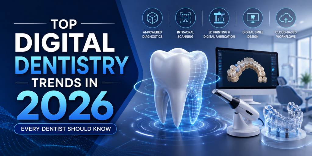
The dental industry is undergoing one of its biggest transformations, driven by rapid advancements in technology. From AI-powered diagnostics to 3D imaging, intraoral scanners, and digital workflows, dental practices are embracing smarter solutions that improve efficiency, accuracy, and patient care. As patient expectations continue to evolve, investing in digital technology is no longer just an option—it has become a necessity for clinics that want to remain competitive.
Today, patients expect faster appointments, painless procedures, and clear communication about their treatment. At the same time, dentists are looking for technologies that reduce manual work, improve diagnostic accuracy, and streamline clinical workflows. This is where digital dentistry is making a remarkable impact.
Whether you’re opening a new clinic or upgrading an existing practice, understanding the latest Digital Dentistry Trends in 2026 can help you make informed decisions and prepare your practice for the future. In this article, we’ll explore the technologies that are reshaping modern dentistry and why they matter for both dentists and patients.
Why Digital Dentistry Is Growing Faster Than Ever
Over the past few years, digital transformation has accelerated across healthcare, and dentistry is no exception. Traditional methods that relied heavily on manual impressions, film-based X-rays, and paper records are gradually being replaced by digital systems that offer greater speed, precision, and convenience.
One of the biggest reasons behind this shift is the increasing demand for accurate diagnosis and predictable treatment outcomes. Digital technologies provide dentists with detailed clinical information, enabling them to make better decisions while reducing the chances of errors. At the same time, patients appreciate shorter appointments, improved comfort, and better visualization of their treatment plans.
Another driving factor is the growing adoption of implant dentistry, clear aligner therapy, cosmetic dentistry, and full-mouth rehabilitation. These treatments require precise planning, making advanced digital equipment an essential part of modern dental practice
Artificial Intelligence Is Changing the Way Dentists Diagnose
Artificial Intelligence (AI) has become one of the most exciting developments in modern dentistry. Rather than replacing dentists, AI works alongside clinicians by analyzing dental images and highlighting potential areas of concern. This technology helps improve diagnostic confidence and supports more informed treatment planning.
Modern AI software can assist in detecting conditions such as dental caries, periodontal bone loss, periapical lesions, impacted teeth, and other abnormalities. By reviewing digital radiographs and CBCT scans, AI can identify subtle patterns that may be difficult to detect with the naked eye.
Benefits of AI in Dentistry
- Faster diagnosis
- Early disease detection
- Improved treatment planning
- Better patient communication
- Reduced human error
CBCT Imaging Is Becoming the Standard for Advanced Diagnosis
Among all the innovations in dental imaging, Cone Beam Computed Tomography (CBCT) has become one of the most significant. Unlike conventional two-dimensional X-rays, a CBCT machine captures detailed three-dimensional images of the teeth, jawbone, nerves, sinuses, and surrounding structures.
This comprehensive view allows dentists to diagnose complex cases with greater accuracy and confidence. CBCT imaging has become indispensable in procedures such as dental implant planning, oral surgery, endodontics, orthodontics, and temporomandibular joint (TMJ) evaluations.
Why dentists are adopting CBCT
- High-resolution 3D imaging
- Better implant planning
- Accurate endodontic diagnosis
- Orthodontic assessment
- Safer surgical procedures
For patients, CBCT also improves communication. Instead of relying on verbal explanations, dentists can use detailed 3D images to demonstrate the condition and explain the recommended treatment, helping patients make informed decisions about their oral health.
Intraoral Scanners Are Replacing Traditional Dental Impressions
Few patients enjoy traditional dental impressions. The discomfort of impression trays and impression materials has long been a challenge in restorative and orthodontic dentistry. This is why Intraoral Scanners (IOS) are becoming one of the most valuable investments for modern dental clinics.
An intraoral scanner captures highly accurate digital impressions within minutes, eliminating the need for conventional impression materials. The result is a faster, cleaner, and more comfortable experience for both the patient and the clinician.
Digital impressions also improve collaboration between dental clinics and laboratories. Since scans can be shared instantly, treatment planning becomes more efficient, reducing turnaround time for crowns, bridges, aligners, and implant restorations.
In addition to improving workflow, intraoral scanners help reduce remakes caused by impression inaccuracies. This not only saves time but also lowers overall treatment costs while enhancing patient satisfaction.
Digital Workflows Are Improving Clinical Efficiency
One of the most impactful changes in modern dentistry is the shift toward fully digital workflows. Instead of relying on multiple disconnected systems, clinics are integrating imaging devices, intraoral scanners, CAD/CAM software, and dental laboratories into a seamless digital ecosystem.
This connected workflow minimizes manual errors, reduces paperwork, and speeds up every stage of treatment—from diagnosis and planning to restoration delivery. Dentists can access patient records, radiographs, and digital impressions in one place, making collaboration easier and more efficient.
Digital workflows are particularly beneficial for implant dentistry, prosthodontics, orthodontics, and cosmetic treatments, where precision and communication play a critical role. By reducing delays and improving coordination, clinics can deliver faster treatment without compromising
Dental Lasers Continue to Transform Soft Tissue Procedures
Laser technology has become increasingly popular for soft tissue treatments due to its precision and minimally invasive approach. Compared to traditional surgical techniques, Dental Lasers often result in less bleeding, reduced postoperative discomfort, and faster healing.
Laser dentistry is commonly used for procedures such as gingivectomy, frenectomy, crown lengthening, periodontal therapy, implant uncovering, and soft tissue contouring. The ability to perform these treatments with greater precision enhances both clinical outcomes and patient comfort.
As more patients seek minimally invasive treatment options, dental lasers are becoming an important addition to modern practices that aim to deliver advanced, patient-centered care.
What Does the Future of Digital Dentistry Look Like?
The future of dentistry is increasingly connected, intelligent, and data-driven. Over the next few years, we can expect greater integration of Artificial Intelligence, cloud-based patient management, enhanced CAD/CAM workflows, advanced imaging technologies, and even robotics in specialized procedures.
These innovations will continue to simplify complex treatments, improve diagnostic accuracy, and create a more personalized patient experience. Clinics that adopt digital solutions today will be better prepared to meet future demands and maintain a competitive edge in an evolving healthcare landscape.
Rather than replacing the expertise of dentists, technology will continue to enhance clinical decision-making and enable better outcomes for patients.
Experience the Future of Digital Dentistry with Vatech India
As digital dentistry continues to transform modern dental practices, choosing the right technology partner is more important than ever. At Vatech India, we are committed to helping dentists deliver accurate diagnoses, efficient workflows, and exceptional patient care through advanced digital imaging and treatment solutions.
Whether you’re looking to upgrade your clinic with CBCT systems, Intraoral Scanners, RVG sensors, or Dental Lasers, our team is here to help you find the right solution for your practice.
Ready to take your clinic to the next level? Contact Vatech India today to schedule a product demonstration, speak with our experts, or learn more about our innovative digital dentistry solutions. Together, let’s build the future of smarter, more efficient dental care.
- Published in Uncategorized
How to Choose the Best CBCT Machine for Your Dental Clinic in 2026
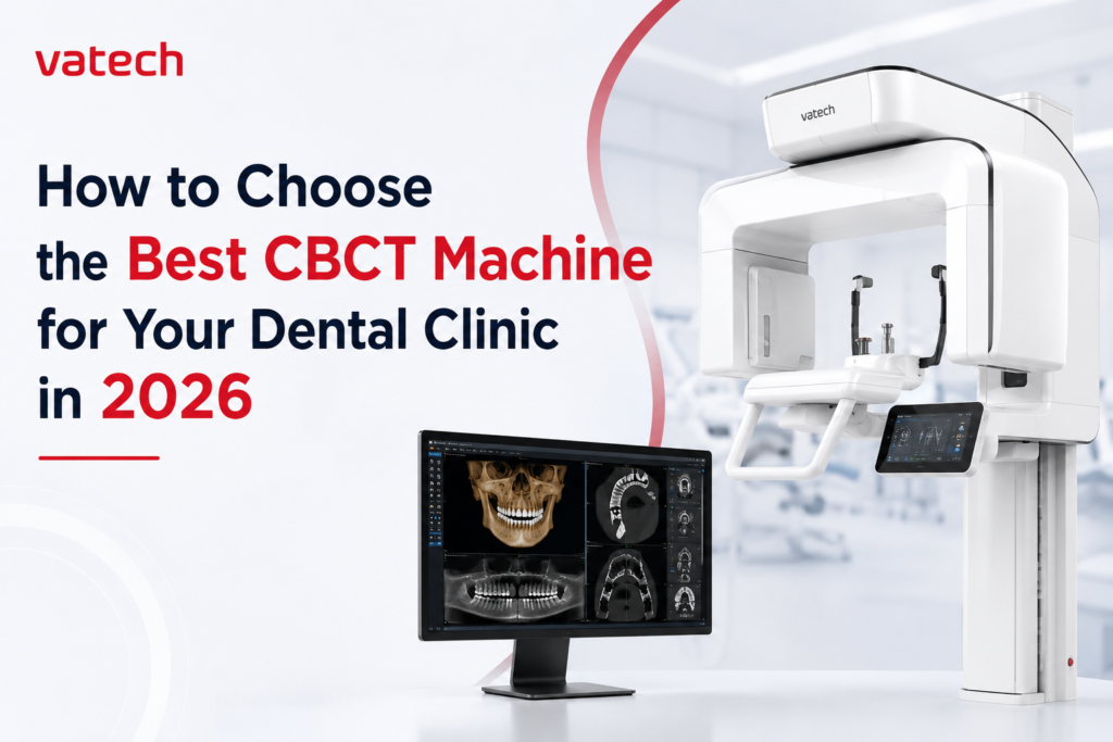
Choosing a CBCT machine for your dental clinic is no longer just about upgrading your equipment—it’s about investing in the future of your practice. As digital dentistry continues to evolve in 2026, dentists are expected to deliver faster diagnoses, more predictable treatment outcomes, and an exceptional patient experience. The right CBCT dental machine helps you achieve all three.
However, with so many brands, models, and technical specifications available in the market, selecting the best CBCT machine can quickly become overwhelming. Manufacturers often promote features such as higher resolution, larger Field of View (FOV), AI-powered software, and lower radiation doses. While these specifications are important, the real question every dentist should ask is: Which CBCT machine is the right fit for my clinic and my patients?
The answer isn’t always the most expensive system or the one with the longest feature list. The best Cone Beam CT machine is one that aligns with your clinical workflow, supports your long-term growth, and delivers consistent diagnostic accuracy.
Whether you’re setting up your first dental clinic, upgrading from a panoramic X-ray system, or expanding your implant and orthodontic services, this guide will help you make an informed decision. By the end of this article, you’ll know exactly what to look for when investing in a CBCT machine for your dental clinic.
Why Choosing the Right CBCT Machine for your Dental Clinic in 2026
The role of dental imaging has changed significantly over the past decade. Modern dentistry is no longer limited to treating visible problems—it focuses on precision, prevention, and patient education. A high-quality CBCT scanner provides detailed three-dimensional images that allow clinicians to visualize anatomical structures far beyond what conventional 2D radiography can offer.
Today, CBCT technology plays a crucial role in:
- Dental implant planning
- Orthodontic treatment planning
- Endodontic diagnosis
- Oral and maxillofacial surgery
- TMJ evaluation
- Airway assessment
- Impacted tooth analysis
- Surgical guide planning
More importantly, patients have become increasingly aware of advanced dental technology. They often associate modern equipment with higher standards of care. When patients see detailed 3D images of their oral condition, they gain a clearer understanding of their treatment needs, which can improve communication and increase treatment acceptance.
In short, investing in the right CBCT machine is not just about better imaging—it’s about building trust, improving efficiency, and supporting the long-term growth of your practice.
Before Comparing Machines, Understand Your Clinic's Needs
One of the biggest mistakes dentists make is comparing CBCT machines before defining their own clinical requirements. A machine that works perfectly for a large implant center may not be the best choice for a general dental practice.
Before evaluating any system, ask yourself a few important questions:
- What treatments do I perform most frequently?
- Am I planning to expand into implantology or orthodontics?
- How many scans will I perform each week?
- Will multiple doctors use the machine?
- Do I want a system that can grow with my practice over the next five to ten years?
Your answers will help determine the type of imaging system that best suits your workflow.
For example, a clinic focused on endodontics may prioritize ultra-high image resolution for detecting fine root fractures and complex canal anatomy. On the other hand, an orthodontic practice may require a larger Field of View to evaluate the entire craniofacial structure.
Choosing a CBCT machine for your dental clinic based on your actual clinical needs prevents unnecessary spending and ensures that every feature you invest in adds value to your practice.
Advantages of In-House 3D Imaging with CBCT
Adopting in house CBCT 3D imaging can provide your practice with numerous advantages. We encourage you to read our special article, The Benefits of In-House 3D Imaging Systems, for a thorough examination of these advantages.
The benefits are difficult to overlook, ranging from improving patient satisfaction to increasing referral rates and raising the value of your practice. CBCT technology can open up new revenue streams for your business by providing a greater variety of treatment alternatives and boosting patient confidence through accurate diagnosis.
Additionally, the market offers a wide variety of CBCT machines, each with special benefits. Purchasing CBCT scanners can result in considerable returns on investment, particularly in situations such as single or two implant surgeries, when a specific number of scans can produce a significant ROI.
CBCT vs. OPG: Which Imaging System Is Right for Your Dental Practice?
When choosing dental imaging equipment, many dentists compare CBCT (Cone Beam Computed Tomography) and OPG (Orthopantomogram). While both play an important role in diagnosis, they are designed for different clinical needs.
Understanding how these two imaging systems differ can help you choose the right technology for your practice and provide the best possible care for your patients.
CBCT: Detailed 3D Imaging for Advanced Dental Procedures
A CBCT machine captures highly detailed three-dimensional images of the teeth, jawbone, nerves, and surrounding anatomical structures. This level of detail allows dentists to diagnose complex cases with greater confidence and accuracy.
Because of its advanced imaging capabilities, CBCT has become the preferred choice for procedures such as dental implant planning, orthodontics, endodontics, oral and maxillofacial surgery, TMJ evaluation, and airway analysis. The ability to view the patient’s anatomy from multiple angles helps clinicians make more informed treatment decisions and reduce the risk of complications.
Which One Should You Choose?
The right choice depends on the type of treatments your clinic offers.
If your practice focuses mainly on routine check-ups and general dentistry, an OPG system may be sufficient for your day-to-day diagnostic needs.
However, if you regularly perform dental implants, complex root canal treatments, orthodontic procedures, or oral surgery, investing in a CBCT machine can significantly improve diagnostic accuracy and treatment planning. Its three-dimensional imaging provides information that simply cannot be captured with traditional panoramic X-rays.
Many modern dental clinics choose systems that combine both CBCT and panoramic imaging, giving them the flexibility to perform routine examinations as well as advanced diagnostics using a single platform. This approach not only improves workflow but also prepares the practice for future growth in digital dentistry.
Don't Overlook After-Sales Service and Technical Support
Purchasing a CBCT machine for your clinic is only the beginning of your journey. What truly determines the long-term value of your investment is the quality of support you receive after installation.
Before making your decision, ask questions such as:
- How quickly can the service team respond to a support request?
- Is technical support available in your region?
- Are genuine spare parts readily available?
- Does the company provide installation and hands-on training?
- Are software updates included?
- What warranty and maintenance options are offered?
Choosing a manufacturer with a strong service network can save both time and operational costs in the long run.
Why After-Sales Support Matters
A CBCT machine is a long-term investment, and consistent support ensures it continues to perform reliably for years. Fast response times, preventive maintenance, and access to experienced service engineers help minimize downtime and keep your practice running smoothly.
Choose a CBCT Machine with Low Radiation Dose
One of the most common concerns patients have about CBCT imaging is radiation exposure. Fortunately, advances in imaging technology have made it possible to achieve exceptional image quality while significantly reducing radiation dose.
Modern CBCT systems incorporate intelligent exposure settings, optimized scan protocols, and selectable Fields of View to ensure that patients receive only the amount of radiation necessary for accurate diagnosis.
When comparing CBCT machines, consider questions such as:
- Does the system offer low-dose imaging modes?
- Can exposure settings be adjusted based on the patient’s age or clinical need?
- Does the machine support small FOV scans to limit unnecessary exposure?
Choosing a CBCT machine that follows the ALARA principle—keeping radiation “As Low As Reasonably Achievable”—demonstrates a commitment to patient safety without compromising diagnostic quality.
Which CBCT Machine Is Best for Your Dental Clinic?
After understanding what makes a great CBCT machine, the next question is obvious: Which CBCT system actually delivers these features?
While every clinic has different requirements, some CBCT systems consistently stand out because of their image quality, software capabilities, reliability, and after-sales support. Here are some of the leading options available in 2026.
VATECH Green X
- Best For: Premium digital dentistry
- Strengths: Excellent image quality, multiple FOV options, AI-powered software, low-dose imaging, and reliable after-sales support.
- Considerations: A premium investment best suited for clinics seeking long-term value and advanced diagnostic capabilities.
VATECH A9
- Best For: High-volume and multi-specialty dental practices
- Strengths: Large Field of View (FOV), advanced diagnostics, and versatile clinical workflows.
- Considerations: Ideal for clinics requiring comprehensive imaging and handling complex cases.
Carestream
- Best For: General dental practices
- Strengths: Reliable imaging performance and a well-established global presence.
- Considerations: Features and capabilities vary depending on the model selected.
Planmeca
- Best For: Large dental practices and advanced clinics
- Strengths: Comprehensive digital ecosystem with integrated imaging and workflow solutions.
- Considerations: Higher overall investment compared to many competing brands.
Dentsply Sirona
- Best For: Clinics looking for integrated digital workflows
- Strengths: Strong software integration and digital treatment planning capabilities.
- Considerations: Premium pricing with a higher overall cost of ownership.
Conclusion: Invest in the Right CBCT Machine for Long-Term Success
A CBCT machine is more than just another piece of equipment—it’s an investment in the future of your dental practice. The right system can improve diagnostic accuracy, streamline workflows, increase patient confidence, and support a wider range of treatments, from routine consultations to complex implant surgeries and orthodontic cases.
While factors such as image quality, Field of View (FOV), software capabilities, radiation dose, and after-sales support are all important, your final decision should be based on the long-term value the system brings to your practice rather than the initial purchase price.
A high-quality CBCT machine can help reduce referrals, improve treatment acceptance, enhance patient communication, and create new opportunities for practice growth. Over time, these benefits contribute to a stronger return on investment (ROI), making the technology an asset rather than an expense.
If you’re looking for a CBCT solution that combines advanced imaging technology, flexible scanning options, intuitive software, and dependable after-sales support, VATECH offers a range of systems designed to meet the needs of modern dental practices. Whether you’re a general dentist, implantologist, orthodontist, or managing a multi-specialty clinic, choosing the right imaging partner today can help your practice stay ahead tomorrow.
Ready to upgrade your clinic with advanced CBCT technology? Explore VATECH’s range of dental imaging solutions and discover how the right investment can support better clinical outcomes, greater efficiency, and long-term practice growth.
- Published in Uncategorized


