
Made for smart dentists
Designed with input from clinicians and diagnosticians to put image quality, ergonomics, ease of use and practicality first.
- CMOSHigh-performance sensor technology
- LowerRadiation dose per exposure
- WiderUsable exposure range
- 1Sensor for the whole mouth
What is actually inside
A radiograph is only ever as good as the thing that captured it. Everything downstream — the software, the screen, the diagnosis — is working with whatever the sensor managed to record in that fraction of a second.
High-performance CMOS
VATECH sensors are built on high-performance CMOS technology. X-rays strike a scintillator layer that converts them to light, that light is guided down to the CMOS array, and the array reads it out as a digital image immediately. There is no plate to carry, no scanner to feed, and no processing step between the exposure and the image.
Direct digital, not scanned
That distinction matters. A phosphor plate system still asks someone to carry a plate to a reader and scan it, which is faster than a darkroom but is still a step. A direct digital sensor removes the step entirely — and removes with it every opportunity for the image to be degraded, mishandled or lost between the chair and the screen.
- X-raysArrive from the tube head and strike the first layer.
- ScintillatorConverts them into light.
- CMOS arrayReads that light out as a digital image, immediately.
- Down the cableNo plate to carry, no scanner to feed, no processing step.
Room to be wrong
Not every exposure lands on the setting you intended. What matters is how far off it can be and still give you an image you can read.
VATECH sensors work extremely well across a broader range of exposures, delivering clinically usable images at a wider range of settings than other sensors — even when radiograph conditions are not ideal. A wider usable band is what fewer retakes actually means in practice: an exposure that would have been discarded on another system is still diagnostic on this one, so the patient is not exposed a second time.
Bands are illustrative of relative behaviour, not measured values; usable range varies by exposure setup, anatomy and clinical requirement.
Less dose for the same diagnosis
Superior visible resolution and contrast in the sensors and software produce unparalleled radiographs that are extremely clear, sharp and highly detailed — all while requiring lower radiation exposure. The sensor recovers more information from less signal, so the exposure needed to reach a diagnostic image is lower to begin with.
There is a second, quieter dose saving too. Every retake is an extra exposure the patient did not need, so a system that gets a usable image the first time is lowering total dose even when its per-exposure figure is unchanged. Consistency and dose are the same conversation.
Bar lengths are relative and illustrative, not measured dose values. Actual exposure depends on the unit, the setting and the anatomy being imaged.
Resolution is not the whole story
Resolution is the number everyone compares, because it is the one that fits on a spec sheet. On its own it says surprisingly little about whether you can see a lesion.
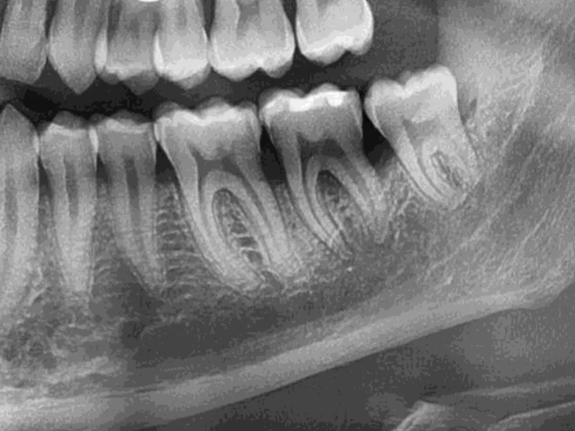
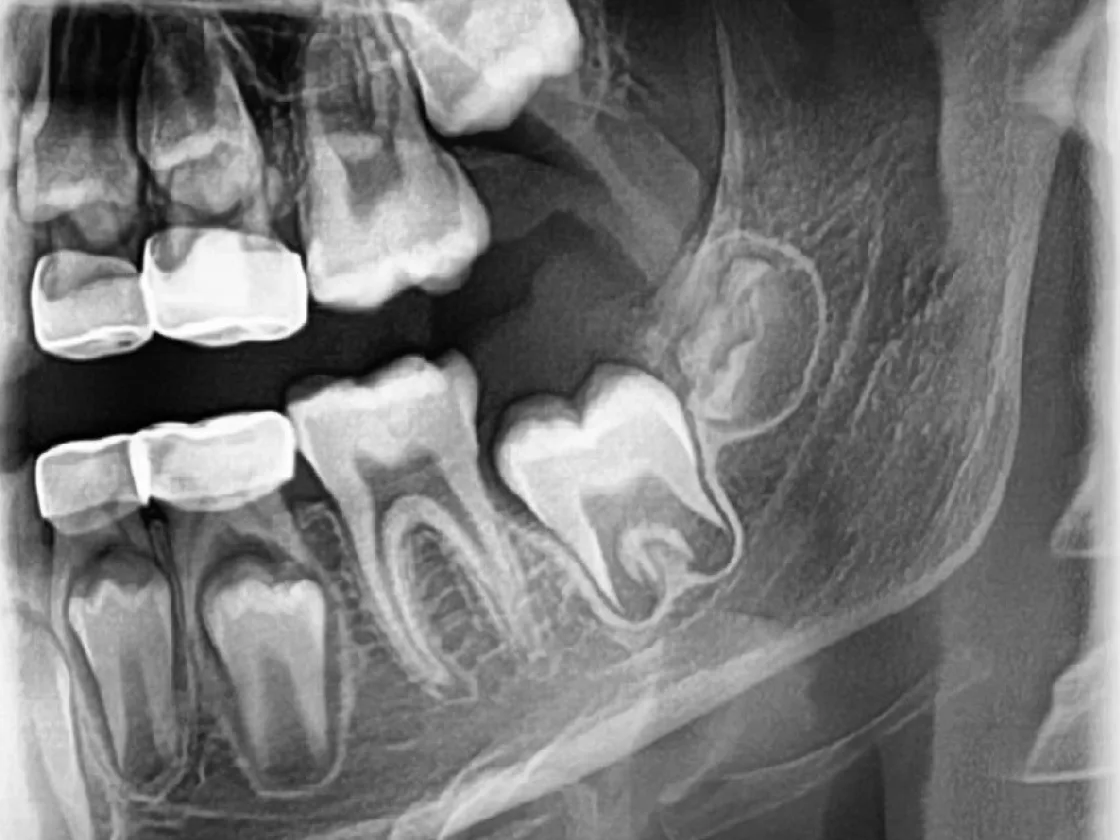
What you actually read a radiograph with is contrast — the separation between one tissue density and the next — and how much noise is sitting on top of it. A high-resolution image with poor contrast separation gives you a very sharp picture of something you still cannot call. VATECH tunes all three together: superior visible resolution and contrast in the sensors and software, producing radiographs that are extremely clear, sharp and highly detailed.
The image-processing presets are part of that, not a cosmetic layer on top of it. Endo, perio and caries each need a different rendering of the same captured data, and having those a click away means the image you read is the one suited to the question you are asking.
Built for a working surgery
A sensor lives a harder life than a spec sheet suggests. It gets handled, wiped, sleeved and dropped.
Sealed housing
Built to be wiped down and barrier-sleeved between every patient.
Cable strain relief
The cable junction is the thing that fails first, so it is reinforced.
Autoclavable holders
Positioning holders that survive a real sterilisation cycle.
USB-powered
No battery to charge, degrade or replace mid-list.
Built for daily handling
Designed for a device that is picked up and put down all day.
Reliable in tough conditions
Performs well even when radiograph conditions are not ideal.
Consistency, dose and retakes are not three separate advantages. They are one property of the sensor, described from three directions — a system that stays diagnostic when the exposure is not perfect asks less of the patient and less of you.
Made for smart dentists.Designed with input from clinicians and diagnosticians, and built to keep working in a real surgery.
Make VATECH your preferred choice
Experience the difference firsthand with a live demonstration at your clinic.